Archive of Biomedical Science and Engineering
Construction and characterization of murine single-chain variable fragment (muscFv) antibody against acrylamide in coffee
Sukanya Ponphimai1, Parichat Srinok1, Nopporn Naewwan1, Thitimakorn Namhong1, Jeeraphong Thonongsaksrikul3, Sanong Suksaweang1,2, Theeraya Simawaranon1,2 and Kanyarat Thueng-In1,2*
2School of Pathology, Institute of Medicine, Suranaree University of Technology, Thailand
3Department of Biomedical Sciences, Faculty of Allied Health Sciences, Thammasat University, Thailand
Cite this as
Ponphimai S, Srinok P, Naewwan N, Namhong T, Thueng-In K, et al. (2024) Construction and characterization of murine single-chain variable fragment (muscFv) antibody against acrylamide in coffee. Arch Biomed Sci Eng 10(1): 009-016. DOI: 10.17352/abse.000032Copyright
© 2024 Ponphimai S, et al. This is an open-access article distributed under the terms of the Creative Commons Attribution License, which permits unrestricted use, distribution, and reproduction in any medium, provided the original author and source are credited.Ingredients of food, especially sugar and starch at high-temperature cooking processes could lead to the formation of acrylamide (AA). This chemical is a harmful carcinogen, a neurotoxicant, a reproductive toxicant, and a carcinogen in animal species. However, the detection of acrylamide contamination in food goes unnoticed. In this work, the mouse monoclonal antibody in the form of a single chain variable fragment (scFv) specific to acrylamide was selected from the murine scFv (muscFv) phage-displayed library. Acrylamide (AA) was used as an antigen for bio-panning. The murine single-chain variable fragment (muscFv) antibody specific to acrylamide in coffee was successfully constructed, which was determined by ELISA and HPLC. Currently, this is the first study, which describes the selection of antibodies against acrylamide from the muscFv phage-displayed library and could be used as a tool for the detection of acrylamide in coffee.
Introduction
Acrylamide (AA), an unsaturated-amide small molecule, is a by-product of food heating processes (due to the Maillard reaction) that are commonly present in cooked foods. This agent was known to be toxic to humans [1,2] It is rapidly absorbed after ingestion and distributed in many organs such as the thymus, liver, heart, brain, and kidneys [3,4]. Due to its genotoxicity and carcinogenicity [5], acrylamide was classified as a Group 2A carcinogen by the International Agency for Research on Cancer (IARC, 1994) and a Category 2 carcinogen and Category 2 mutagen by the European Union.
Acrylamide contamination in food is first reported by the Swedish National Food Administration using the LC/MS/MS method. The most common foods often found containing acrylamide are potato chips, french fries, baked bread, chocolate, and coffee [6]. Many researchers have confirmed the presence of acrylamide in different processed foods. For Thai foods, acrylamide has been found in curries, commercial and conventional snacks, instant noodles as well as coffee that contain starch and fat as the major components and are cooked at high temperatures [7]. In addition, as reported in the EFSA’s scientific opinion on AA in food, the exposure data reported that coffee was one of the sources of this toxicant in adult diets. Several methods for acrylamide detection are reported in the literature, including high-performance liquid chromatography (HPLC) [8], gas chromatography [9], gas chromatography coupled with mass spectrometry (GC-MS) [10], liquid chromatography coupled with mass spectrometry (LC-MS/MS) [11,12], immunoassay [13] electro-chemical- biosensors [14] , fluorescent method [4] and quartz microbalance sensors [15] . Most of the methods need advanced equipment. This is an important issue because acrylamide is suspected to cause cancer in humans and there is no concern about acrylamide contaminated [16]. Thus, a device for detecting acrylamide in foods is urgently required. In this study, the muscFv specific to acrylamide was selected and tested against acrylamide using enzyme-linked immunosorbent assay (ELISA).
Materials and methods
Antibodies and reagent
Mouse anti-M13, goat anti-mouse HRP-conjugated polyclonal antibody (GE Healthcare, Dako, Denmark, EU), Mouse monoclonal anti-C-Myc (BioLegend, San Diego, USA), T4 DNA Ligase, NcoI and Notl restriction enzymes (Thermo Scientific, USA), Acrylamide standard (AA) and Isopropyl-β-D-thio-galacto-pyranoside (IPTG) (Sigma-Aldrich, USA) pSEX81, pOPE101 phagemid vector (Progen Biotechnik GmbH, Heidelberg, Germany) were used in this study.
Selection of murine scFv specific to acrylamide
The murine single chain variable fragment (muscFv) phage display library was kindly provided by Asst. Prof. Dr. Jeeraphong Thanongsaksrikul from the Faculty of Allied Health Sciences, Thammasat University, Thailand [17]. The AA was used as the antigen to select single-chain fragments (scFv) from the library by bio-panning [17]. After bio-panning, XL-1 Blue E.coli colonies containing recombinant muscFv-pSEX81 surface expression phagemid vector (Progen Biotechnik GmbH, Heidelberg, Germany) were tested by direct colony PCR using pelB Primer 5›-ATACCTATTGCCTAC GGCAGC-3› and gIII Primer 5›-TAGCATTCCACAG ACAGCCC-3›. The muscFv display phage particles were rescued from each individual E.coli clone by co-infecting the bacteria with M13KO7 helper phages (GE Healthcare Life Sciences, Denmark, EU). The titers of the rescued phages were determined and normalized for acrylamide-specific binding testing by indirect ELISA. Acrylamide and BSA were used as antigens and blank, respectively. The E.coli clone containing muscFv that gave optical density at 450 nm. two times higher than blank were selected and subcloned into the pOPE101- plasmid (Progen Biotechnik GmbH, Heidelberg, Germany) for recombinant murine scFv production.
Small-scale muscFv expression
The XL-1 Blue E.coli containing muscFv-pOPE101 phagemid vector was grown in 10 mL LB-broth supplemented with 100 μg/mL ampicillin in a 37 °C shaker until OD600 nm = 0.6 Isopropyl-β-D-thio-galacto-pyranoside (IPTG) (Sigma-Aldrich, USA) was added to a final concentration of 1 mM. Cells were harvested 3 h later, and centrifuged at 10,000 rpm for 20 min at 4 °C. The cells were lysed by sonication in a lysis buffer.
Binding analysis by indirect ELISA
The E.coli lysate was tested for binding to acrylamide standard (AA) according to the protocols described [17]. Briefly, acrylamide standard (5 µg) and BSA in 100 μl coating buffer (0.05 M carbonate-bicarbonate buffer, pH 9.6) were immobilized on ELISA wells and kept at 37 °C until dry. After washing with PBST, all wells were blocked with 3% BSA in PBS and kept at 37°C for 1 h. Next, the standardized E.coli lysate was individually added into wells and kept at room temperature for 1 h. After washing, mouse monoclonal anti-c-Myc (BioLegend, San Diego, USA) diluted 1:3,000 in PBST was added and kept at room temperature for 1 h. The horse-radish-peroxidase conjugated goat anti-mouse (GE Healthcare, Dako, Denmark, EU) (diluted 1:5000 in PBST) was added and kept for 1 h. After washing, ABTS (2,2’-Azinobis [3-ethylbenzothiazo-line-6-sulfonicacid-diammonium salt) chromogenic substrate (KPL. Inc, USA) was added. The absorbance OD at 405 nm was measured. The E.coli lysate clone which gave OD signals two times higher than control was selected for the next experiment.
Expression and purification
The selected XL-1 Blue E.coli transformed with muscFv-pOPE101 were grown in 100 mL LB-broth supplemented with 100 μg /mL ampicillin incubated at 37 °C with shaking. The protein expression was induced by Isopropyl-β-D-thiogalacto-pyranoside (IPTG) (Sigma-Aldrich, a final concentration of 1 mM. Cells were harvested 3 h later, centrifuged at 10,000 rpm for 20 min at 4 °C, and lysed by sonication in lysis buffer (50 mM NaH2PO4, 300 mM NaCl, 10 mM imidazole, pH 8). The E.coli lysate was collected and subjected to Ni-NTA resin (Thermo Scientific, USA) affinity chromatography purification system according to the manufacturer›s instructions. The purified anti-AA muscFv protein was aliquoted and stored at -20 °C until use. The purity of purified muscFv was determined by SDS-PAGE and Coomassie staining.
DNA characterization and sequences
DNA sequences from selected anti-AA muscFv clones were sequenced using pelB (5›-ATACCTATTG-CCTACGGCAGC-3›) and pOPE101 (5› -TAGATCTTCTTCTGAGATCAGC -3›) primers. The determining regions (CDRs) and immunoglobulin framework regions (FRs) of muscFv sequences were analyzed using (IMGT/V-QUEST tool of the International ImMunoGeneTics Information System (IMGT®) [18]. The DNA sequence of anti-AA muscFv was multiple aligned using Bio-Edit and ClustalW2 program.
Homology modeling and intermolecular docking
Anti-AA muscFv sequences were aligned using Bio-Edit and the ClustalW2 program. The position of VH, VL, and Linker according to the vector pOPE101 was determined for validation of protein structure and translated to amino acid via the website (https://-web.expasy.org/translat). Anti-AA muscFv submitted the sequence of each protein to be modeled using the website (https://colab.research.google.com/github/sokrypton/ColabFold/blob/main/Alpha-Fold2.ipynb). The best match model of each protein (antigen and antibody (scFv)) was submitted for docking. The acrylamide 3D structure modeled pdb files for docking assembly were searched (https://www.rcsb.-org/). Docking results were obtained according to their binding affinities by using AutoDock Vina. PyMOL. Discovery Studio was used for the largest docking clusters of the interactive residues, and those with the lowest local energy were selected.
Determination of the cutoff value of Anti-AA muscFv antibody by indirect ELISA
To establish the cutoff value of the indirect ELISA, 10-fold dilutions of the AA standard were used. Each dilution was tested in triplicate. The OD405 nm value plus three times the standard deviation (SD) was used as the cutoff [19]. All experimental samples were considered positive if the OD405 nm value was higher than this cutoff value.
Determination of Anti-AA muscFv specific to acrylamide in coffee
The validity of Anti-AA muscFv specific to acrylamide in coffee was conducted by using 2 brands of dark roasted coffee beans; designated coffee A and coffee B. The spike (0.1, 0.3, 0.5, and 0.7 mg/mL) and non-spike with AA standard in coffee was determined by ELISA as protocol previously described above. Furthermore, the presence of acrylamide in coffee was also confirmed by High-performance liquid chromatography (HPLC).
Results
Selection of anti-AA muscFv antibody specific to acrylamide
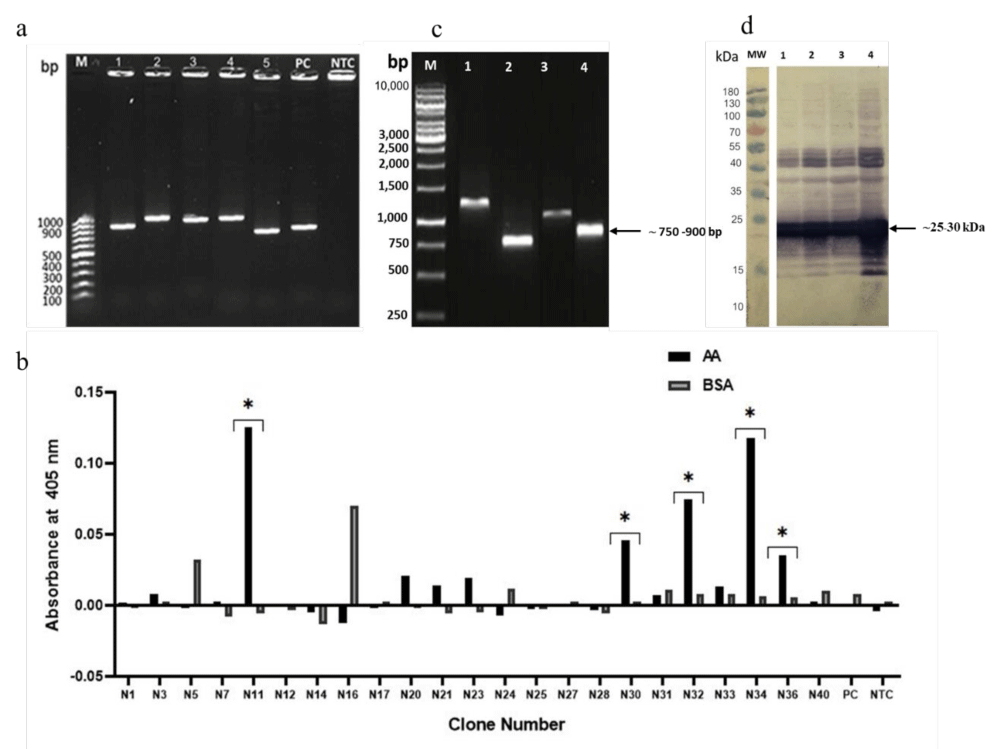
After bio-panning, the E.coli colony containing recombinant phagemid muscFv vector was individually picked for direct colony PCR. Five clones (21.7%) presented positive PCR bands at expected size; ∼750 - 900 bp as shown in Figure 1a. All positive clones were selected for protein expression. All 5 clones (100%) could be expressed muscFv at molecular size ∼25-30 kDa. The ELISA binding activity of these 5 clones is shown in Figure 1b. According to sequencing results and ELISA binding, clone number 11 was selected for sub-clone into the pOPE101 vector for large-scale recombinant protein expression and purification. Colony-directed PCR screening showed four positive PCR amplicons (N11.1, N11.2, N11.3, and N11.4) from 7 colonies screening (71.4%) Figure 1c Protein expression of all 4 clones showed the positive band at the expected molecular mass (∼25 - 30 kDa) as shown in Figure 1d. All 4 clones could bind to acrylamide by ELISA binding result (data not shown).
Expression and purification of anti-AA muscFv antibodies
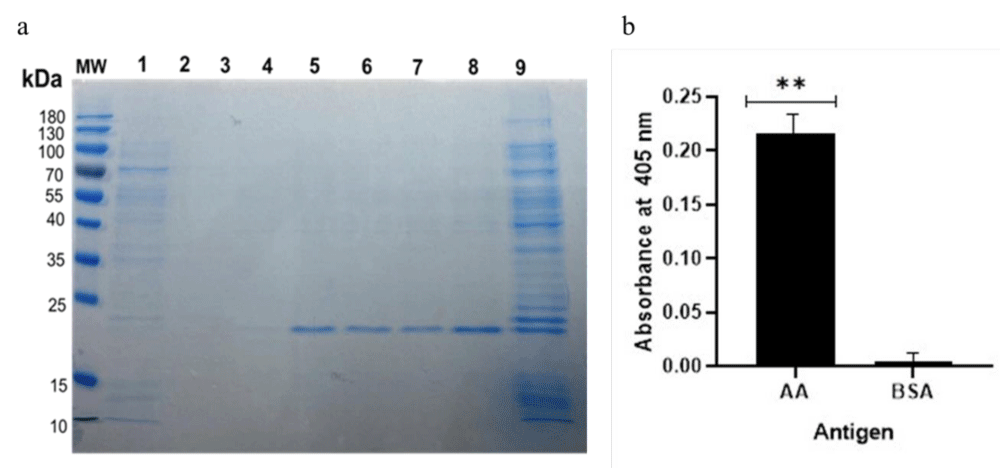
After sub-cloning into the pOPE101 vector, direct colony PCR was performed. The positive PCR at ~750 - 900 bp was selected for protein expression. Clone number 11.1 (MscFv-N11.1) was selected. Figure 2a showed Coomassie staining of the purified muscFv-N11.1 protein.
Purified anti-AA muscFv antibodies bound specifically to acrylamide
The binding activity of purified muscFv-N11.1 was verified via indirect ELISA. Coated AA and BSA wells were incubated sequentially with muscFv-N11.1, mouse monoclonal anti-c-My, HRP-conjugated goat anti-mouse IgG, and ABTS substrate, respectively. The absorbance OD at 405 nm was shown in Figure 2b. The result showed that muscFv-N11.1 could bind specifically to AA (p < 0.001) when compared to BSA.
Characterization and homology modeling and intermolecular docking
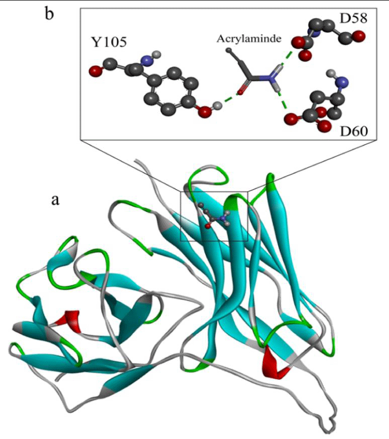
The sequence analysis showed a complete CDR1-3 and immunoglobulin framework of mouse VH and VL as shown in Figure 3. This should be indicated that muscFv-N11.1 was successfully constructed. The molecular docking model of muscFv-N11.1 was performed. Ribbon display model showing muscFv-N11.1 in blue, green, gray, and red. Superimposed picture of the 3D structures of muscFv-N11.1 and acrylamide structures as shown in Figure 4a. muscFv-N11.1 used D-58, D-60, and Y-105 of VH domain to interact with acrylamide via hydrogen bond (energy rang - 12.89 kcal/mol) as shown Figure 4b).
Validation of AA standard and anti-AA muscFv antibody
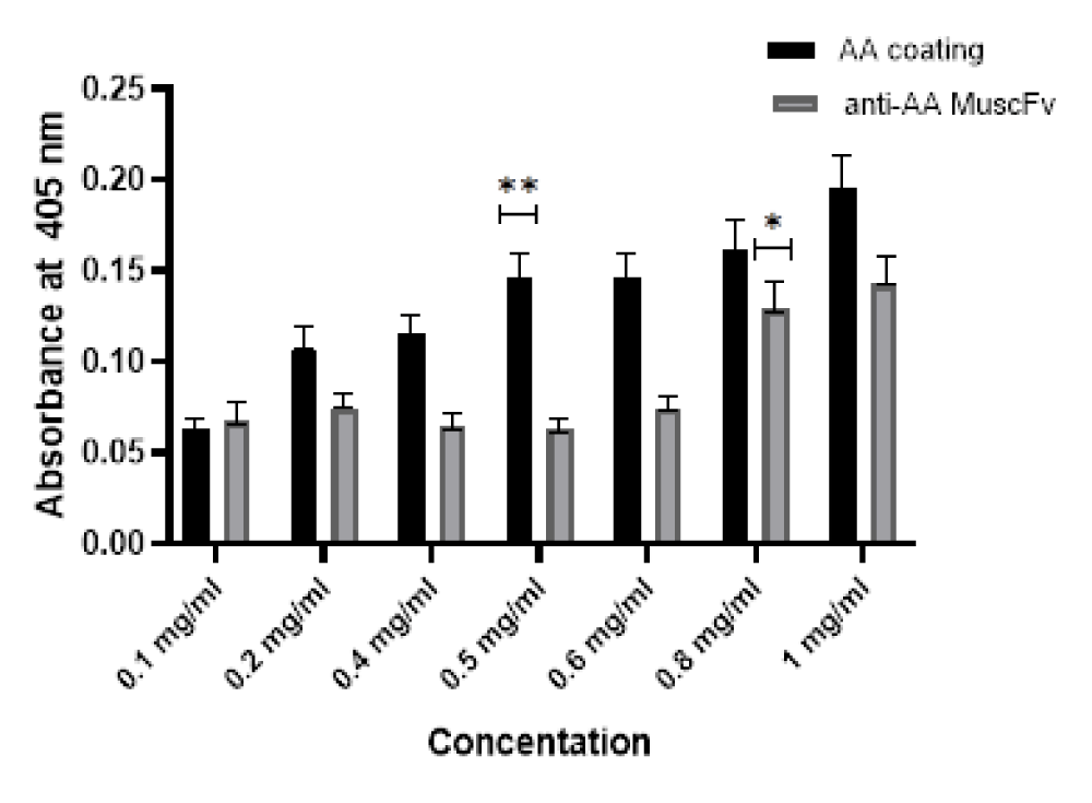
The checker broad titration was used to determine the lowest amount of acrylamide detected (LOD) and the lowest amount of Anti-AA muscFv antibody used in the ELISA. The AA standard was diluted in coating buffer; 0, 0.1, 0.2, 0.4, 0.5, 0.6, 0.8, and 1 mg/mL and coated in each ELISA well. A concentration of 0.5 mg/mL was found to be the lowest concentration that could be detected and gave similar OD405 as the concentration 0.6 and 0.8 mg/mL, respectively. Whereas, the lowest amount of Anti-AA muscFv antibody was 0.8 mg/mL (Figure 5). Triplicate wells of each concentration were performed.
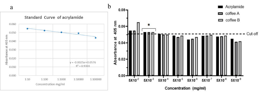
After the acrylamide standard›s test linearity plot. Figure 6a illustrates the linear correlation between concentration and area under the peak was found to be linear across the measured concentration range, with an R2 = 0.9303. To establish the cutoff value of the ELISA, the AA standard was 10-folded diluted, and analyzed. The mean of the OD405 nm values for these samples, as detected by the indirect ELISA, was 0.0510, with a standard deviation of 0.0003, which was calculated using the formula: mean of the negative sample values plus three standard deviations (SDs) [19] (Deshpande For a 99% confidence interval, the cutoff was defined as follows: mean of the negative serum OD405 nm values plus three standard deviations = 0.0510 + 3 × 0.0003 = 0.0519. The sensitivity of this in-house ELISA was 0.005 mg/mL as shown in Figure 6b. Spike-and-recovery and linearity-of-dilution experiments are important methods for validating and assessing the accuracy of ELISA [20]. The concentration of the positive control sample (known concentration) was within the linear section of the standard curve in order to obtain valid and accurate results (data not shown).
Determination of AA muscFv specific for acrylamide in coffee
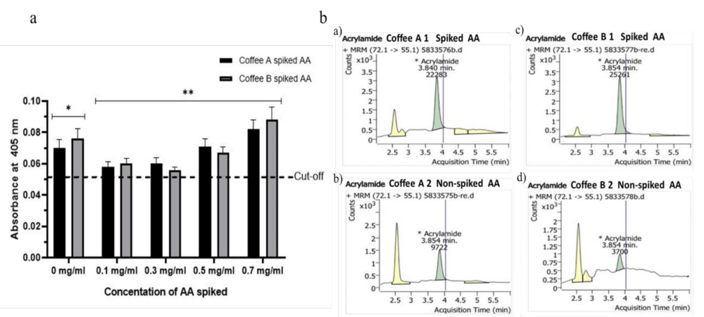
The efficacy of muscFv-N11.1 specific to acrylamide in coffee was performed by ELISA. Two bands of coffee; coffee A and coffee B were randomly selected. The muscFv-N11.1 could detect acrylamide in both spiked and non-spiked coffee as shown in Figure 7a. The bar graph shows an increasing trend that varies with the concentration of AA standard (**). For coffee A and coffee B 0 mg/mL served as the negative controls (NC) (*) were able to detect acrylamide in coffee A and coffee B at similar concentrations (0.5 and 0.7 mg/mL). Each experiment was performed in triplicate. Values were presented as the means ± SD. All experimental samples› values were higher than the cutoff value (0.0519). The result was also confirmed by HPLC that both bands of coffee have acrylamide in Figure 7b. The spiked AA in coffee showed an OD405 increase when the AA spiked concentration increased, which was shown in Figure 7a. The result revealed a dose-dependent fashion. Thus, the produced muscFv-N11.1 could bind specifically to acrylamide.
Discussion
Acrylamide belongs to a group of toxins that is harmful to human and animal health, due to both acute and chronic effects, including neurotoxicity, genotoxicity, carcinogenicity, reproductive toxicity, hepatotoxicity, and immunotoxicity [21]. The presence of acrylamide was found predominantly in heat-treated carbohydrate-rich foods, such as potatoes, biscuits, cereals, and coffee; announced by the Swedish National Food Administration in April 2002 [22]. The European Food Safety Authority (EFSA) has determined an estimated dosage range within which the substance is likely to cause potential health effects. Acrylamide has been estimated to range from 425 for average adult consumers down to 50 for high consuming toddlers. In coffee determined it to 400 µg /kg for roasted coffee and not more than 850 µg /kg for instant coffee [23]. Among a variety of established methodologies for analyzing acrylamide, namely high-performance liquid chromatography (HPLC) [8], gas chromatography coupled with mass spectrometry (GC-MS) [9,10] and immunoassay [13] etc. were described. The immunological methods are among the most rapid, simplest, cheapest, and most suitable for on-lab screening.
ELISA, a rapid method based on the recognition of antigen-antibody binding with high specificity and affinity, utilizes optical detection of colored products catalyzed by enzyme labels. Due to its specificity and affinity, coupled with efficient enzymatic catalysis, ELISA methods offer optimal recovery and adaptability for detecting AA in various food samples, including Chocolate products, Cocoa powder, Nuts, French fries, roast potatoes, potato crisps/chips, biscuits, baked, and toasted [21]. The advantages such as affordability, simplicity, ease of handling, and portability, prove particularly effective in identifying AA in thermally processed foods [24]. ELISA also demonstrates good sensitivity, selectivity, high-throughput capabilities, and compatibility with other technologies like biotin-avidin amplification and chemiluminescence, thus attracting increased attention for AA detection in foods [25]. However, the multiple washing and incubation steps in ELISA extend the detection process, making it more suitable for primary screening of food products with excessively high AA concentrations during processing or on the market. In comparison, standard methods like LC-MS/MS and GC-MS provide rapid detection, meeting the needs of food industries, regulatory bodies, and consumers [26]. Nevertheless, they require further refinement to enhance accuracy, sensitivity, repeatability, reproducibility, multi-step, and portability for achieving online and real-time detection of trace amounts of AA [27].
In this study, mouse variable heavy and light chain formed of muscFv-N11.1 was selected from the muscFv-phage library. The muscFv -phage library size utilized in this experiment with substantial antibody sequence diversity, which was comparable to the other non-immune libraries previously reported [28,29]. Similarly, in a related study using an immunized phage display library a specific nanobody termed Nb-7E against an acrylamide derivative xanthyl acrylamide (XAA) was isolated from an immunized phage display library and confirmed to be able to detect acrylamide [25]. muscFv-N11.1 was found to be specific to acrylamide by using established ELISA. The result confirmed by HPLC that both bands of coffee have acrylamide. The chromatograms showed acrylamide peak and interference corresponding to an acrylamide calibration standard with a retention time of 3.8 min. Similarly, a study in-depth study of acrylamide formation in coffee during roasting: role of sucrose decomposition and lipid oxidation using determination of acrylamide in coffee by liquid chromatography-tandem mass spectrometry. Acrylamide peak showed a retention time of 2-3.1 min [30]. Corresponding to the study determination of acrylamide after Its extraction from potato chips was acrylamide spiked in chips sample and analyzed using ultrasound-assisted liquid-liquid extraction (UA-LLE) technique / HPLC-UV showed acrylamide peak at a retention time of 4.15 min [31]. Computerized docking models showed that muscFv-N11.1 used the VH domain (D-58, D-60, and Y-105) to interact with acrylamide via hydrogen bonds. The binding efficacy of muscFv-N11.1 to acrylamide was less than 0.005 mg/mL.
Conclusion
In this study, the selection of antibodies against acrylamide from murine scFv phage-displayed library was performed in order to select used by acrylamide standard: AA and N-acryloxy succinimide: NAS used as antigen for bio-panning were expressed and purified to be specific for acrylamide contaminated in food. Anti-AA muscFv against acrylamide was produced, and an indirect ELISA was developed for the determination of coffee. The application to samples from coffee indicated that ELISA may be used for the estimation of total acrylamide concentrations. The method has shown satisfactory results in terms of specificity and accuracy, which were confirmed by High-Performance Liquid Chromatography (HPLC).
We would like to thank the study participants and all others who helped directly or indirectly to accomplish this study. Especially, we would like to thank the Department of Translational Medicine, Institute of Medicine, Suranaree University of Technology. We thank Jeeraphong Thonongsaksrikul for providing the muscFv phage-displayed library. And we thank Kanyarat Thueng-in for consulting in the lab.
Ethics statement
This study protocol was reviewed and approved by the Biosafety Control Board at Suranaree University of Technology, Nakhon Ratchasima, Thailand (SUT-IBC-03/2021).
Author contributions
SP performed most experiments and drafted the manuscript. JT carried out the selection of the phage library against acrylamide and contributed to the genetic, molecular, and immune biochemical characterization of muscFv. In addition, SP participated in the design and coordination of the study. PS and NN performed some of the experiments such as purification and bio-panning procedures for muscFv isolation and selection. TN performed some of the experiments such as designing ELISA for binding testing. SS, TS, and KTI helped to consult in the lab and draft the manuscript. Furthermore, KTI participated in the design and coordination of the entire research project. All authors have read and approved the final version of the manuscript.
Funding
This work was supported by research grants of Graduate Studies from the Office of the National Research Council of Thailand for the year 2021 (Grant number D-M6111380-1/2564).
- Tareke E, Rydberg P, Karlsson P, Eriksson S, Törnqvist M. Acrylamide: a cooking carcinogen? Chem Res Toxicol. 2000 Jun;13(6):517-22. doi: 10.1021/tx9901938. PMID: 10858325.
- Svensson K, Abramsson L, Becker W, Glynn A, Hellenäs KE, Lind Y, Rosén J. Dietary intake of acrylamide in Sweden. Food Chem Toxicol. 2003 Nov;41(11):1581-6. doi: 10.1016/s0278-6915(03)00188-1. PMID: 12963011.
- Capuano E, Fogliano V. Acrylamide and 5-hydroxymethylfurfural (HMF): A review on metabolism, toxicity, occurrence in food and mitigation strategies. LWT - Food Science and Technology. 2011; 44:793–810. https://doi.org/10.1016/j.lwt.2010.11.002
- Hu Q, Xu X, Li Z, Zhang Y, Wang J, Fu Y, Li Y. Detection of acrylamide in potato chips using a fluorescent sensing method based on acrylamide polymerization-induced distance increase between quantum dots. Biosens Bioelectron. 2014 Apr 15;54:64-71. doi: 10.1016/j.bios.2013.10.046. Epub 2013 Oct 31. PMID: 24252761.
- Xu Y, Cui B, Ran R, Liu Y, Chen H, Kai G, Shi J. Risk assessment, formation, and mitigation of dietary acrylamide: current status and future prospects. Food Chem Toxicol. 2014 Jul;69:1-12. doi: 10.1016/j.fct.2014.03.037. Epub 2014 Apr 5. PMID: 24713263.
- Rosén J, Hellenäs KE. Analysis of acrylamide in cooked foods by liquid chromatography tandem mass spectrometry. Analyst. 2002 Jul;127(7):880-2. doi: 10.1039/b204938d. PMID: 12173642.
- Komthong P, Suriyaphan O, Charoenpanich J. Determination of acrylamide in Thai-conventional snacks from Nong Mon market, Chonburi using GC-MS technique. Food Addit Contam Part B Surveill. 2012;5(1):20-8. doi: 10.1080/19393210.2012.656145. Epub 2012 Feb 27. PMID: 24779691.
- Gökmen V, Senyuva HZ, Acar J, Sarioğlu K. Determination of acrylamide in potato chips and crisps by high-performance liquid chromatography. J Chromatogr A. 2005 Sep 23;1088(1-2):193-9. doi: 10.1016/j.chroma.2004.10.094. PMID: 16130751.
- Shin D-C, Kim C-T, Lee Y-C. Reduction of acrylamide by taurine in aqueous and potato chip model systems. Food Research International. 2010; 43:1356–1360. https://doi.org/10.1016/j.foodres.2010.03.024
- Lee MR, Chang LY, Dou J. Determination of acrylamide in food by solid-phase microextraction coupled to gas chromatography-positive chemical ionization tandem mass spectrometry. Anal Chim Acta. 2007 Jan 16;582(1):19-23. doi: 10.1016/j.aca.2006.08.042. Epub 2006 Aug 26. PMID: 17386469.
- Zhang Y, Ren Y, Jiao J, Li D, Zhang Y. Ultra high-performance liquid chromatography-tandem mass spectrometry for the simultaneous analysis of asparagine, sugars, and acrylamide in Maillard reactions. Anal Chem. 2011 May 1;83(9):3297-304. doi: 10.1021/ac1029538. Epub 2011 Apr 4. PMID: 21462916.
- Yang LX, Zhang GX, Yang LX, He Y. LC-MS/MS determination of acrylamide in instant noodles from supermarkets in the Hebei province of China. Food Addit Contam Part B Surveill. 2012;5(2):100-4. doi: 10.1080/19393210.2012.658874. Epub 2012 Mar 16. PMID: 24779738.
- Zhou S, Zhang C, Wang D, Zhao M. Antigen synthetic strategy and immunoassay development for detection of acrylamide in foods. Analyst. 2008 Jul;133(7):903-9. doi: 10.1039/b716526a. Epub 2008 Apr 3. PMID: 18575643.
- Batra B, Lata S, Sharma M, Pundir CS. An acrylamide biosensor based on immobilization of hemoglobin onto multiwalled carbon nanotube/copper nanoparticles/polyaniline hybrid film. Anal Biochem. 2013 Feb 15;433(2):210-7. doi: 10.1016/j.ab.2012.10.026. Epub 2012 Oct 24. PMID: 23103399.
- Kleefisch G, Kreutz C, Bargon J. Quartz Microbalance Sensor for the Detection of Acrylamide. Sensors. 2004; 4:136–146. https://doi.org/10.3390/s40900136
- Kumar J, Das S, Teoh SL. Dietary Acrylamide and the Risks of Developing Cancer: Facts to Ponder. Front Nutr. 2018 Feb 28;5:14. doi: 10.3389/fnut.2018.00014. PMID: 29541638; PMCID: PMC5835509.
- Thanongsaksrikul J, Srimanote P, Tongtawe P, Glab-Ampai K, Malik AA, Supasorn O, Chiawwit P, Poovorawan Y, Chaicumpa W. Identification and production of mouse scFv to specific epitope of enterovirus-71 virion protein-2 (VP2). Arch Virol. 2018 May;163(5):1141-1152. doi: 10.1007/s00705-018-3731-z. Epub 2018 Jan 22. PMID: 29356992.
- Brochet X, Lefranc MP, Giudicelli V. IMGT/V-QUEST: the highly customized and integrated system for IG and TR standardized V-J and V-D-J sequence analysis. Nucleic Acids Res. 2008 Jul 1;36(Web Server issue):W503-8. doi: 10.1093/nar/gkn316. Epub 2008 May 24. PMID: 18503082; PMCID: PMC2447746.
- Deshpande SS Enzyme Immunoassay from Concept to Product Development, 1st ed. Kluwer Academic Publishers, New York. 1996.
- Lequin RM. Enzyme immunoassay (EIA)/enzyme-linked immunosorbent assay (ELISA). Clin Chem. 2005 Dec;51(12):2415-8. doi: 10.1373/clinchem.2005.051532. Epub 2005 Sep 22. PMID: 16179424.
- Rifai L, Saleh FA. A Review on Acrylamide in Food: Occurrence, Toxicity, and Mitigation Strategies. Int J Toxicol. 2020 Mar/Apr;39(2):93-102. doi: 10.1177/1091581820902405. Epub 2020 Feb 4. PMID: 32013673.
- Taeymans D, Wood J, Ashby P, Blank I, Studer A, Stadler RH, Gondé P, Van Eijck P, Lalljie S, Lingnert H, Lindblom M, Matissek R, Müller D, Tallmadge D, O'Brien J, Thompson S, Silvani D, Whitmore T. A review of acrylamide: an industry perspective on research, analysis, formation, and control. Crit Rev Food Sci Nutr. 2004;44(5):323-47. doi: 10.1080/10408690490478082. PMID: 15540646.
- European Food Safety Authority (EFSA); Benford D, Bignami M, Chipman JK, Ramos Bordajandi L. Assessment of the genotoxicity of acrylamide. EFSA J. 2022 May 5;20(5):e07293. doi: 10.2903/j.efsa.2022.7293. PMID: 35540797; PMCID: PMC9069548.
- Quan Y, Chen M, Zhan Y, Zhang G. Development of an enhanced chemiluminescence ELISA for the rapid detection of acrylamide in food products. J Agric Food Chem. 2011 Jul 13;59(13):6895-9. doi: 10.1021/jf200954w. Epub 2011 Jun 14. PMID: 21639145.
- Liang Y, Zeng Y, Luo L, Xu Z, Shen Y, Wang H, Hammock BD. Detection of Acrylamide in Foodstuffs by Nanobody-Based Immunoassays. J Agric Food Chem. 2022 Jul 27;70(29):9179-9186. doi: 10.1021/acs.jafc.2c01872. Epub 2022 Jul 12. PMID: 35819336; PMCID: PMC10111249.
- Desmarchelier A, Hamel J, Delatour T. Sources of overestimation in the analysis of acrylamide-in coffee by liquid chromatography mass spectrometry. J Chromatogr A. 2020 Jan 11;1610:460566. doi: 10.1016/j.chroma.2019.460566. Epub 2019 Sep 23. PMID: 31582163.
- Aykas DP, Urtubia A, Wong K, Ren L, López-Lira C, Rodriguez-Saona LE. Screening of Acrylamide of Par-Fried Frozen French Fries Using Portable FT-IR Spectroscopy. Molecules. 2022 Feb 9;27(4):1161. doi: 10.3390/molecules27041161. PMID: 35208950; PMCID: PMC8879073.
- Thanongsaksrikul J, Srimanote P, Maneewatch S, Choowongkomon K, Tapchaisri P, Makino SI, Kurazono H, Chaicumpa W. A V H H that neutralizes the zinc metalloproteinase activity of botulinum neurotoxin type A. J Biol Chem. 2010 Mar 26;285(13):9657-9666. doi: 10.1074/jbc.M109.073163. Epub 2010 Jan 21. PMID: 20093370; PMCID: PMC2843216.
- Thattanon P, Thanongsaksrikul J, Petvises S, Nathalang O. Monoclonal antibody specific to the Dia blood group antigen generated by phage display technology. Blood Transfus. 2020 Sep;18(5):366-373. doi: 10.2450/2020.0031-20. Epub 2020 Jul 22. PMID: 32931415; PMCID: PMC7592165.
- Kocadağlı T, Göncüoğlu N, Hamzalıoğlu A, Gökmen V. In depth study of acrylamide formation in coffee during roasting: role of sucrose decomposition and lipid oxidation. Food Funct. 2012 Sep;3(9):970-5. doi: 10.1039/c2fo30038a. Epub 2012 Jul 16. PMID: 22796869.
- Ghalebi M, Hamidi S, Nemati M. High-Performance Liquid Chromatography Determination of Acrylamide after Its Extraction from Potato Chips. Pharmaceutical Sciences. 2019; 25:338–344. https://doi.org/10.15171/PS.2019.42
Article Alerts
Subscribe to our articles alerts and stay tuned.
 This work is licensed under a Creative Commons Attribution 4.0 International License.



 Save to Mendeley
